|
Plate V.
Click image to enlarge
 |
|
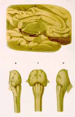
Plate III. Charcot's Original Lesion Characterization:
Fig. A. Left cerebral hemisphere, medial aspect. "Sclerosed plaques occupying
the corpus callosum" and other parts (a,a) of the ventricle wall (CC, Corpus
callosum; CH, hippocampal gyrus; CO, thalamus).
Figg. B,C,D. "Sclerosed plaques" on spinal bulb and pons (dark, olive-green
areas), as seen from the right side (B), left side (D), and anteriorly (C).
Distinctive Pathological Findings:
Fig. A. The section severing the two cerebral hemispheres shows an injury
having coherently surged up, in a series of partly peaked, partly rounded
waves, off of the corpus callosum undersurface into its substance. Cerebral
cortex and paraventricular nuclei (grey matter compartments!) have not been
spared.
|
|
Fig. A for the first time shows lesion waves and spikes invading the corpus
callosum off of its underface.
Figg. B and D demonstrate the specific lateral spinal cord patches' mode
of upper termination. Noteworthy, as to fig. C, are the two thin
lesion-streaks extending lengthwise, in the lower spinal bulb, down the midst
of the pyramids. In addition, figg. B,C, D all show many compact plaques
scattered over the pons,
|
|
In figg. B and D, there appear specific scars' strictly aligned with the
spinal cord's flanks (cf. Plate I; Plate II, fig. 1; Plate II; Plate IV, fig.
3), looking as though they had been "torn out" from, in particular, the
uppermost part of their extent. Fig. C shows a comparable involvement of the
pyramids.
Significance:
Presented in Charcot's "Lectures", 1884 edition, these "unpublished findings"
impressively illustrate in what singular way cerebral multiple sclerosis
spreads.
|