|
Plate VI.
Click image to enlarge
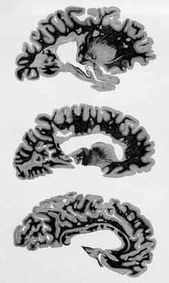
Special Lesion Characteristics:
In addition to a projection of a number of compact and many finely dentate, basally connected lesions off of the ventricular border, the cerebral hemisphere's three slices parallel to the median plane display a gradual lesion detachment and eventual free scattering outwards of the first involved zone into the entire cerebrum.
|
|
Siemerling and Raecke's Original Description:
The cerebral hemisphere's paramedian sections show "extensive plaques, strongly involving, even penetrating the corpus callosum" (lower figure); "many others affect both cerebral white matter and cortex; the most strongly sclerosed, i.e. scarred, areas are in the ventricle walls" .
Otherwise, the account on the fourth case of Siemerling and Raecke's 1914 report merely mentioned that also spinal cord, brain stem, cerebellum, and optical nerves had at all levels been comparably involved.
Significance:
In this plate, the ventricle-based lesion picture appears as a sort of intermediate between the damages projected off of the ventricular border, shown in Plate V, fig. A, and the vast epiventricular lesion depicted in Plate IV, fig. 1. Illustrated for the first time here is that specific cerebral plaques may also emerge, apparently independently, peripheral to the ventricle-based lesions. Besides, it is once more shown that the grey matter, though poor in myelin, is not spared.
|