|
Plate VIII.
Click image to enlarge
 |
|
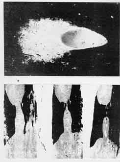
Fig. A. Scheinker's Original Cues: In 1947, this picture was said to
illustrate how the emergence of the multiple sclerosis plaques is related to
the vascular system or, more specifically, to veins (117). In 1954, the injury
was merely commented on as a "Small area of myelin loss in the vicinity of a
distended and congested small blood vessel." (118)
Fig. B. Original Description by Steiner in 1962:
Three longitudinal transverse sections through the posterior part of a lower
thoracic spinal cord of a "classical case of multiple sclerosis" (hindmost
section on the left, frontmost section on the right side).
The arrows in the two left figures' midsts indicate a bridging between upper
and lower posterior patch along the spinal cord's posterior median
septum [increasing towards the frontmost section]. The most anterior
transverse section (figure on the right side), shows very irregular
[variegated and bizarre] damages to the cord's sides (lateral
arrows).
|
|
Specific Lesion Traits:
The injury, burst forth to only the one side of a vein's circumference, looks
as though the tissue had, on the part of this vessel, been splashed apart and
aside. This first illustration of a specific cerebral plaque's dynamic
expansion from its vein has remained unmatched as to its expressiveness.
|
|
Specific Findings:
The frontwardly increasing injury appears most specifically characterized by
the saw-toothed damages to the intermediate section's left side as well as the
rhomboid lesions along the frontmost section's right and flame-shaped patches
in its left side -- all of which can hardly be understood otherwise than as
effects of tensile impacts.
Documentary Significance:
The splashing of a specific cerebral plaque from the one side of a vein (fig.
A) and the serration and crenelation of the damages to the spinal cord's sides
(fig. B) indicate that these lesions are primarily attributable to mechanical
forces.
|