|
Plate XII.
Click image to enlarge
 |
Ā |
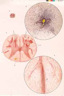
Eichhorst's 1913 Observations on Spinal Cord Lesions Caused by a
Blood-borne Agent (Smallpox-Virus):
The spinal cord cross-sections show, both at a weak (fig. 1) and stronger
(fig. 2) magnification, that the "inflammatory foci's" points and
stripes(densely dotted) are marked by heavy perivascular
infiltrates of mostly mononuclear cells (fig. 3), red blood cells (fig. 2, B)
or both sorts of cells (fig. 2, E,E).
Fig. 2: B, compact hemorrhage; E,E, "inflammatory foci".
Fig. 4: sld, "inflammatory infiltrate" seaming a blood vessel of the
spinal cord's posterior median septum.
|
Characteristics of a Systemically Disseminated Spinal Cord Affection:
In the macroscopically inconspicuous spinal cord, microscopically a great
number of strictly perivascular lesions, up to the size of a millet seed,
appear spread through especially the spinal cord's central grey matter. In the
lower spinal cord, the white matter is also involved. But of these white matter
lesions, most connect with foci in the spinal cord's central grey matter --
only a minor part lying entirely within the white substance, and even fewer
lesions reaching the spinal cord's circumference. Overall, the lesions appear
so irregularly scattered that their pattern of spread is, even on closely
adjacent spinal cord cross-sections, hardly ever comparable.
|
|
Relevance of the Documentation: Although this was not the first time that
the picture of a "disseminated myelitis", i.e. multifocal spinal cord injury
originating in a blood-borne agent's dispersion into the spinal cord was
illustrated, Eichhorst's picture characterized this kind of injury particularly
aptly. Regarding the septa-related injuries of multiple sclerosis, his image of
a conformable "inflammatory septum affection" caused by a blood vessel's course
within the latter (fig. 4, sld) deserves particular attention.
|